Anti Lamin Immunofluorescence

Lamin B1 Antibody 119d5 F1 Nuclear Envelope Marker Ab8982 Abcam

Alexa Fluor 647 Anti Lamin B1 Antibody Epr8985 B Ab194108 Abcam

Alexa Fluor 488 Anti Lamin B2 Antibody Epr9701 B Ab200426 Abcam

Anti Lamin A Antibody Ko Tested Ab26300 Abcam

Alexa Fluor 594 Anti Lamin A Lamin C Antibody Epr4100 Ab215324 Abcam

Alexa Fluor 647 Anti Lamin B2 Antibody Epr9701 B Ab200427 Abcam
Additionally anti lamin a c terminal antibodies are suitable for use in immunoblotting approx.
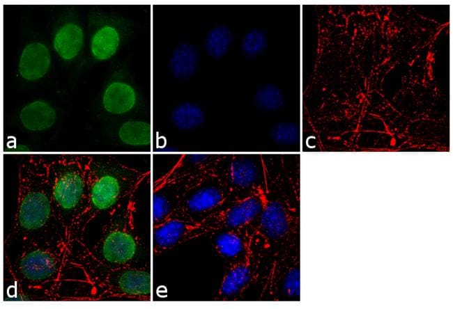
Anti lamin immunofluorescence. The cells were methanol fixed 5 min and incubated with the antibody ab16048 1µg ml for 1h at room temperature. Supplied as 100 µg purified antibody 0 5 mg ml. Antibody dilution 1 1 000 2. The cells were fixed with 4 paraformaldehyde for 10 minutes permeabilized with 0 1 triton x 100 for 10 minutes and blocked with 1 bsa for 1 hour at room temperature.
Immunocytochemistry immunofluorescence anti lamin a antibody 133a2 ab8980 these images show hela cells stained with laminin a antibody ab8980 green and with dapi blue. Lamin a antibody ma1 06101 in if immunofluorescence analysis of lamin a was performed using 70 confluent log phase hela cells. Anti lamin a c terminal antibody is suitable for use in western blot 0 1 0 2 μg ml using hela nuclear extract. Invitrogen anti lamin b1 monoclonal l 5 catalog 33 2000.
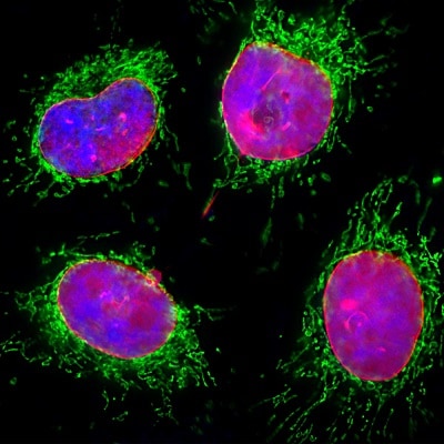
The anti lamin a c antibody reveals strong nuclear lamina staining while anti lamp1 antibody reveals strong cytoplasmic punctate staining of lysosomes and early endosomes. This antibody can also be used in indirect immunofluorescence 1 2 μg ml using hela cells rat nrk and mouse 3t3 cells. Since both dna blue and lamin a c red are associated with the nuclear compartment this region appears crimson in this image. Tested in western blot wb immunofluorescence if immunoprecipitation ip and elisa elisa applications.
Laminin from engelbreth holm swarm murine sarcoma basement membrane was separated on sds page and probed with rabbit anti laminin ab11575. Specificity of the anti laminin antibodies is determined by indirect immunofluorescent labeling of formalin fixed paraffin embedded human or animal tissue sections and by dot blot immunoassay. The cleavage of lamins results in nuclear dysregulation and cell death 5 6. During apoptosis lamin a c is specifically cleaved into a large 41 50 kda and a small 28 kda fragment 3 4.
The antibody was developed using goat anti rabbit igg peroxidase secondary antibody and a chemiluminescent substrate. Lamin a c is cleaved by caspase 6 and serves as a marker for caspase 6 activation. This antibody reacts with chicken human mouse rat samples. For immunofluorescence analysis hela cells were fixed and permeabilized for detection of endogenous lamin b1 using anti lamin b1 recombinant rabbit monoclonal antibody product 702972 1 100 dilution and labeled with goat anti rabbit igg h l superclonal secondary antibody alexa fluor 488 conjugate product a27034 1 2000.
By indirect immunofluorescence the antibody demonstrates specific basement membrane staining of enzymatically unmasked human and animal tissue. 2000 j struct biol 129 313 23.

7497 Phospho Ndrg1 Thr346 D98g11 Xp Rabbit Mab Alexa Fluor 647 Conjugate Cst抗体 Confocal Immunofluorescent Analysis Of Mab Painting Alexa

Alexa Fluor 488 Anti Lamin A Lamin C Antibody Ep4520 16 Ab205769 Abcam

Recombinant Anti Lamin B1 Antibody Epr22165 121 Bsa And Azide Free Ko Tested Ab239399

Recombinant Anti Lamin B1 Antibody Epr8985 B Ko Tested Ab133741 Abcam

Anti Lamin A Lamin C Antibody Wl4g10 Ko Tested Ab232730 Abcam

Alexa Fluor 488 Anti Lamin A Lamin C Antibody Epr4100 Ab185014 Abcam
Lamin B1 Antibody Pa5 19468

Anti Lamin A Lamin C Antibody Wl4g10 Bsa And Azide Free Ko Tested Ab269575
Anti Lamin A Lamin C Antibody 4c4 Ko Tested Ab190380 Abcam

Alexa Fluor 647 Anti Lamin B Receptor Lbr Antibody E398l Ab201349 Abcam

Anti Laminin 1 2 Antibody Ab7463 Abcam

Anti Lamin A Lamin C Antibody 4c11 Bsa And Azide Free Ko Tested Ab244577

Anti Lamin B1 Antibody Nuclear Envelope Marker Ko Tested Ab16048 Abcam

